Fungal Infections

November 19, 2008

Valley Fever – Coccidiodomycosis

Valley Fever – Coccidiodomycosis

Definition

Valley fever is caused by fungi in the soil. The fungi that cause valley fever can be stirred into the air by anything that disrupts the soil, such as farming, construction and wind. The fungi can then be breathed into the lungs. Valley fever is a form of coccidioidomycosis (kok-sid-e-oi-doh-mi-KOH-sis), or cocci (KOK-si) infection. It can cause fever, chest pain and coughing, among other signs and symptoms.

More than half of those who inhale the valley fever fungi have few, if any, problems. But some, especially pregnant women, people with weakened immune systems, and those of Asian, Hispanic and African descent, may develop a more serious and sometimes fatal form of coccidioidomycosis infection.

Mild cases of valley fever usually go away on their own. In more severe coccidioidomycosis infections, doctors prescribe antifungal medications that can treat the underlying infection.

Symptoms
Valley fever is one of three forms of coccidioidomycosis. The forms include: acute (valley fever), chronic, and disseminated.

Acute coccidioidomycosis (valley fever)

The acute form is often mild, with few, if any, symptoms. When signs and symptoms do occur, they appear one to three weeks after exposure. They tend to resemble those of the flu, and can range from minor to severe:

Fever
Cough
Chest pain — varying from a mild feeling of constriction to intense pressure resembling a heart attack
Chills
Night sweats
Headache
Fatigue
Shortness of breath
Joint aches
Red, spotty rash
The rash that sometimes accompanies valley fever is made up of painful red bumps that may later turn brown. The rash mainly appears on your lower legs but sometimes on your chest, arms and back. Others may have a raised red rash with blisters or eruptions that look like pimples.

If you don’t become ill from valley fever, you may only learn that you’ve been infected when you later have a positive skin or blood test. Small areas of residual infection (nodules) in the lungs that show up on a routine chest X-ray may also be found. Although the nodules typically don’t cause problems, they can look like cancer on X-ray, leading to biopsies.

If you do develop symptoms, especially severe ones, the course of the disease is highly variable. It can take from six months to a year to fully recover, and fatigue and joint aches can last even longer. The severity of the disease depends on several factors, including your overall health and the number of fungus spores you inhale.

Chronic coccidioidomycosis

Appearing as many as 20 years after the initial infection, chronic pneumonia due to coccidioidomycosis is most common in people with diabetes or weakened immune systems. You’re likely to have periods of worsening symptoms alternating with periods of recovery. Signs and symptoms are similar to those of tuberculosis:

Low-grade fever
Weight loss
Cough
Chest pain
Blood-tinged sputum
Nodules in the lungs

Disseminated coccidioidomycosis

The most serious form of the disease, disseminated coccidioidomycosis occurs when the infection spreads (disseminates) beyond the lungs to other parts of the body. Most often these parts include the skin, bones, liver, brain, heart, and the membranes that protect the brain and spinal cord (meninges).

The signs and symptoms of disseminated disease depend on which parts of your body are affected and may include:

Nodules, ulcers and skin lesions that are more serious than the rash that sometimes occurs with other forms of the disease
Painful lesions in the skull, spine or other bones
Painful, swollen joints, especially in the knees or ankles
Meningitis — an infection of the membranes and fluid surrounding the brain and spinal cord and the most deadly complication of valley fever

Causes

The fungi that cause valley fever — Coccidioides immitis or Coccidioides posadasii — thrive in the alkaline desert soils of southern Arizona, northern Mexico and California’s San Joaquin Valley. They’re also endemic to New Mexico, Texas and parts of Central and South America — areas with mild winters and arid summers.

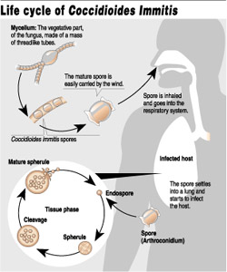
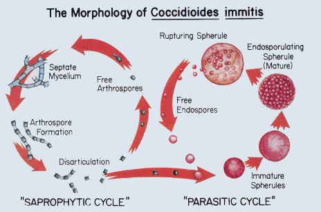
Like many other fungi, coccidioides species have a complex life cycle. In the soil, they grow as a mold with long filaments that break off into airborne spores when the soil is disturbed. The spores are extremely small, can be carried hundreds of miles by the wind and are highly contagious. Once inside the lungs, the spores reproduce, perpetuating the cycle of the disease.

Risk factors

Environmental exposure. Anyone who inhales the spores that cause valley fever is at risk of infection. Some experts estimate that up to half the people living in areas where valley fever is common have had the disease. People who have jobs that expose them to dust are most at risk — construction, road and agricultural workers, ranchers, archeologists, and military personnel on field exercises.

Smoking. Smokers, especially those with scarring and thickening of lung tissue (pulmonary fibrosis), are at higher risk of valley fever and subsequent chronic pneumonia than are nonsmokers.

Race. For reasons that aren’t well understood, Filipinos, Hispanics, blacks and Asians are more susceptible to developing serious infection with coccidioidomycosis than are whites.

Pregnancy. Pregnant women are vulnerable to more serious coccidioidomycosis in the third trimester and right after their babies are born.

Diabetes. Valley fever infection may be more severe among people with diabetes.
Weakened immune system. Anyone with a weakened immune system is at increased risk of serious complications, including disseminated disease. This includes people living with AIDS or those being treated with steroids, chemotherapy or anti-rejection drugs after transplant surgery. People with cancer and Hodgkin’s disease also have an increased risk.

Age. Older adults are more likely to develop valley fever than younger people are. This may be because their immune systems are less robust or because they have other medical conditions that affect their overall health.

When to seek medical advice

Valley fever, even when it’s symptomatic, often clears on its own. Yet for older adults and others at high risk, recovery can be slow and the risk of disseminated disease high. For that reason, it’s important to seek medical care if you develop the signs and symptoms of valley fever, especially if you live in or have recently traveled to an area where this disease is common.

Be sure to tell your doctor if you’ve traveled to a place where valley fever is endemic and have symptoms. More and more, people who spend a few days golfing or hiking in Arizona return home with valley fever but are never tested for the disease.

Tests and diagnosis

Valley fever isn’t diagnosed on the basis of signs and symptoms, which are usually vague and nonspecific, or on a chest X-ray, which can’t distinguish valley fever from other lung diseases. Instead, a definitive diagnosis depends on finding Coccidioides spherules (cysts) in tissue, blood or other body secretions. For that reason, you’re likely to have one or more of the following tests:

Sputum smear or culture. These tests check a sample of your sputum for the presence of the valley fever fungus.

Blood tests. Through a blood test, your doctor can check for antibodies against the fungus that causes valley fever.

Complications

Valley fever can cause a number of serious complications, especially in people living with HIV/AIDS.

These complications include:

Severe pneumonia. Most people recover from valley fever pneumonia without complications. Others, mainly Filipinos, Hispanics, blacks and Asians and those with weakened immune systems, may become seriously ill.

Ruptured lung nodules. A small percentage of people develop thin-walled nodules (cavities) in their lungs. Many of these eventually disappear without causing any problems, but some may rupture, causing chest pain and difficulty breathing. A ruptured lung nodule might require the placement of a tube into the space around the lungs to remove the air, or surgery to repair the damage.
Disseminated disease. This is the most serious complication of coccidioidomycosis. If the fungus spreads (disseminates) throughout the body, it can cause problems ranging from skin ulcers and abscesses to bone lesions, severe joint pain, heart inflammation, urinary tract problems and meningitis — an infection of the membranes and fluid covering the brain and spinal cord.

Treatments and drugs

Rest often the only treatment

Most people with acute valley fever don’t require treatment. Even when symptoms are severe, the best therapy for otherwise healthy adults is often bed rest and fluids — the same approach used for colds and the flu. Still, doctors carefully monitor people with valley fever.

Antifungal medications

If symptoms don’t improve or become worse, or if you are at increased risk of complications, your doctor may prescribe an antifungal medication such as fluconazole. Antifungal medications are also used for high-risk people or for those with chronic or disseminated disease.

In general, the antifungal drugs fluconazole and itraconazole are used for all but the most serious cases. All antifungals can have serious side effects. However, these side effects usually go away once the medication is stopped. The most common side effects of fluconazole and itraconazole are nausea, vomiting, abdominal pain and diarrhea. More serious infection may be treated initially with an intravenous antifungal medication such as amphotericin.

These medications control the fungus but sometimes don’t destroy it, and relapses may occur. For many people, a single bout of valley fever results in lifelong immunity, but the disease can be reactivated or you can be reinfected if your immune system is significantly weakened.

Prevention

If you live in or visit areas where valley fever is common, take common-sense precautions, especially during the summer months when the chance of infection is highest. Consider wearing a mask, staying inside during dust storms, wetting the soil before digging, and keeping doors and windows tightly closed.

Mayo Clinic

Valley Fever or Coccidioidomycosis

The technical name for Valley Fever is Coccidioidomycosis, or “Cocci” for short. It is caused by Coddidioides immitis, a fungus somewhat like yeast or mildew which lives in the soil. The tiny seeds, or spores, become wind-borne and are inhaled into the lungs, where the infection starts. Valley Fever is not contagious from person to person. It appears that after one exposure, the body develops immunity.

Valley Fever is a sickness of degree. About 60 percent of the people who breathe the spores do not get sick at all. For some, it may feel like a cold or flu. For those sick enough to go to the doctor, it can be serious, with pneumonia-like symtoms, requiring medication and bed rest.

Of all the people infected with Valley Fever, one or more out of 200 will develop the disseminated form, which is devastating, and can be fatal. These are the cases in which the disease spreads beyond the lungs through the bloodstream – typically to the skin, bones, and the membranes surrounding the brain, causing meningitis.

If you live in an endemic area, you may have had Valley Fever without even knowing it. In some endemic areas, it is estimated that as much as half of the population has been infected. Persons whose activities put them in much contact with the soil appear to have a somewhat greater risk. Once infected, persons of African, Filipino and some other Asian ancestries seem to be at a greater risk of contracting the more serious, or disseminated, form of the disease. The young, the old, and those with lowered immune systems are also in the high risk group. While men are at greater risk than women, pregnant women are especially vulnerable, particularly in the third trimester.

Diagnosis

Usually, diagnosis is made on the basis of one or more of the following three tests: recovery of the Cocci organisms from sputum or some other body fluid; blood tests that reflect the body’s reaction to the presence of the fungus; and skin tests. These tests are quite reliable, but they may fluctuate according to the stage of the disease. Chest X-rays reveal some of the abnormalities associated with Cocci, but the shadows are difficult to distinguish from those of tuberculosis or some other lung disease.

Treatment

Patients suffering from the flu-like symptoms of Cocci will probably be sent to bed by the doctor. Since most cases are mild and self-limiting, there is no consensus on how aggressively it should be treated. However, once serious symptoms appear – including pneumonia and labored breathing – treatment should be prompt. Unfortunately, all four antifungal drugs in use are disagreeable and often toxic. Although it is the “gold standard” of treatment, Amphotericin B is the worst, according to patients, especially to those who have to have it injected beneath the base of their skull for meningitis. Its side effects include nausea, fever and kidney damage. Oral drugs include ketoconazole and more recently, fluconazole and itraconazole.

In severe cases, where the fungus has permanently damaged lung or bone tissue, surgery may be needed. Since the drugs serve only to suppress the fungus, not to kill it, those who develop a severe case of Valley Fever may require treatment for years.

ValleyFever.com

valleyfeverdist1


valleyfeverlifecycle

valleyfeverlifecycle2

Histoplasmosis

>What is histoplasmosis?

histoplasmosis1
Histoplasmosis is a disease caused by the fungus Histoplasma capsulatum. Its symptoms vary greatly, but the disease primarily affect the lungs. Occasionally, other organs are affected. This form of the disease is called disseminated histoplasmosis, and it can be fatal if untreated.

Can anyone get histoplasmosis?

Yes. Positive histoplasmin skin tests occur in as many as 80% of the people living in areas where H. capsulatum is common, such as the eastern and central United States. Infants, young children, and older persons, in particular those with chronic lung disease are at increased risk for severe disease. Disseminated disease is more frequently seen in people with cancer, AIDS or other forms of immunosuppression.

How is someone infected with H. capsulatum?

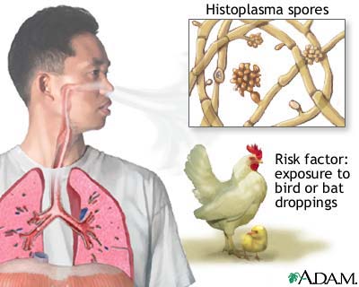
H. capsulatum grows in soil and material contaminated with bat or bird droppings. Spores become airborne when contaminated soil is disturbed. Breathing the spores causes infection. The disease is not transmitted from an infected person to someone else.

What are the symptoms of histoplasmosis?

Most infected persons have no apparent ill effects. The acute respiratory disease is characterized by respiratory symptoms, a general ill feeling, fever, chest pains, and a dry or nonproductive cough. Distinct patterns may be seen on a chest x-ray. Chronic lung disease resembles tuberculosis and can worsen over months or years. The disseminated form is fatal unless treated.

When do symptoms start?

If symptoms occur, they will start within 3 to 17 days after exposure; the average is 10 days.

Is histoplasmosis treatable?

Yes. Antifungal medications are used to treat severe cases of acute histoplasmosis and all cases of chronic and disseminated disease. Mild disease usually resolves without treatment. Past infection results in partial protection against ill effects if reinfected.

Where is H. capsulatum found?

H. capsulatum is found throughout the world and is endemic in certain areas of the United States. The fungus has been found in poultry house litter, caves, areas harboring bats, and in bird roosts.

What can be done to prevent histoplasmosis?

It is not practical to test or decontaminate most sites that may be contaminated with H. capsulatum, but the following precautions can be taken to reduce a person’s risk of exposure:

Avoid areas that may harbor the fungus, e.g., accumulations of bird or bat droppings.

Centers for Disease Control and Prevention

hisstoplasmosis2

Histoplasmosis

Definition

Every year, hundreds of thousands of people worldwide get a lung disease called histoplasmosis. It’s transmitted through airborne spores that you breathe into your lungs when you work in or around soil that contains a fungus called Histoplasma capsulatum. Farmers, landscapers, construction workers and people who have contact with bird or bat droppings are especially at risk for histoplasmosis.

Most people with histoplasmosis never develop signs and symptoms and aren’t aware they have the infection. But for some people — primarily infants and those with compromised immune systems — histoplasmosis can be serious.

Effective treatments are available for even the most severe forms of histoplasmosis. But these therapies often involve extensive hospital stays and can cause serious side effects. That’s why it’s important for people with compromised immune systems to avoid exposure to histoplasmosis.

Symptoms

Several tpes of histoplasmosis exist, ranging from mild to life-threatening. The most benign form produces no signs or symptoms, but severe infections can cause serious problems throughout your body as well as in your lungs. When signs and symptoms do occur, they usually appear three to 17 days after exposure.

Mild to moderate cases

Asymptomatic primary histoplasmosis. This is the most common form of histoplasmosis and usually causes no signs or symptoms in otherwise healthy people who become infected. The only sign of infection may be small scars in the lungs. In that case, special radiologic testing can usually confirm that nodules aren’t cancerous.

Acute symptomatic pulmonary histoplasmosis.

This form of histoplasmosis tends to occur in otherwise healthy people who have had intense exposure to H. capsulatum. Because the severity of the disease depends on the number of fungus spores inhaled, reactions may range from a brief period of not feeling well to serious illness. Typical signs and symptoms include:

Fever
Headache
Dry cough
Chills
Chest pain
Weight loss
Sweats

In some cases, arthritis or pericarditis — an inflammation of the sac that surrounds the heart — may develop weeks or months after the initial infection. These problems aren’t a sign that the infection has spread outside your lungs. Instead, they develop because your immune system responds to the fungus with an unusual amount of inflammation.

On the other end of the spectrum, people who have inhaled a large number of spores may develop severe acute pulmonary syndrome, a potentially life-threatening condition in which breathing becomes difficult. Acute pulmonary syndrome is frequently referred to as spelunker’s lung because it often occurs after intense exposure to bat excrement stirred up by explorers in caves.

Moderate to severe

Chronic pulmonary histoplasmosis. This type of histoplasmosis usually affects people with an underlying lung disease such as emphysema. It’s most common in white, middle-aged men. The disease is chronic and if left untreated may progress to disabling lung problems. Signs and symptoms include:

Fatigue
Fever
Night sweats
A cough that may bring up blood

Disseminated histoplasmosis.

Occurring primarily in infants and people with compromised immune systems, disseminated histoplasmosis can affect nearly any part of the body, including your eyes, liver, bone marrow, skin, adrenal glands and intestinal tract. Untreated disseminated histoplasmosis is usually fatal. Depending on which organs are affected, people with this form of the disease may develop:

Anemia
Pneumonia
Pericarditis
Meningitis
Adrenal insufficiency
Ulcers of the mouth, tongue or intestinal tract

Causes

Histoplasma capsulatum is primarily found in the temperate regions of the world and is the most common fungus in the United States. It’s endemic in the Ohio, Missouri and Mississippi river valleys, where the great majority of people have been exposed.

The fungus thrives in damp soil that’s rich in organic material, especially the droppings from birds and bats. For that reason, it’s particularly common in chicken and pigeon coops, old barns, caves and parks.

Birds themselves aren’t infected with histoplasmosis — their body temperature is too high — but they can carry H. capsulatum on their feathers, and their droppings support the growth of the fungus. Birds commonly kept as pets, such as canaries and parakeets, aren’t affected. And although bats, which have a lower body temperature, can be infected, you can’t get histoplasmosis from a bat or from another person.

Instead, you develop histoplasmosis when you inhale the reproductive cells (spores) of the fungus. The spores are extremely light and float into the air when dirt or other contaminated material is disturbed. That’s why a high number of cases occur in farmers, landscapers, construction workers, spelunkers and people living near construction sites.

Histoplasmosis and your lungs

Because the spores of H. capsulatum are microscopic in size, they can easily enter your lungs and settle in the small air sacs. There, the spores are trapped by macrophages — immune system cells that attack foreign organisms. The macrophages carry the spores to lymph nodes in your chest, where they continue to multiply. This may lead to inflammation, scarring and calcium deposits. In cases of heavy infection, the lymph nodes may become so enlarged that they obstruct your esophagus or your lungs’ airways.

Most often, however, you’re not likely to have noticeable signs and symptoms, and the infection clears on its own without treatment. But if your immune system isn’t able to eliminate the spores, they can enter your bloodstream and travel to other parts of your body. In that case, you may develop a variety of severe problems that can be fatal if not diagnosed and treated quickly.

Risk factors

Anyone exposed to H. capsulatum is likely to become infected. People who inhale a huge number of spores — those who work with heavily infected soil or have close contact with bats, for example — are more likely to develop signs and symptoms.

Most at risk of infection

Farmers
Poultry keepers, especially when cleaning chicken coops, pigeon roosts, and bat-infested barns or lofts
Construction workers, especially those who work around old buildings with roosting birds
Landscapers and gardeners
People involved in building roads
People who monitor bird populations or who have contact with bats or bat caves
Archeologists
Geologists

Most at risk of severe infection

Because their immune systems are weakened, the following people are most likely to develop disseminated histoplasmosis, the more serious form of the disease:

Infants and very young children.
Older adults. The risk of disseminated histoplasmosis increases with age.
HIV-positive people or those with AIDS.

People receiving chemotherapy or long-term treatment with corticosteroid drugs such as prednisone.
People who have had organ transplants and are taking anti-rejection medications.

When to seek medical advice

Contact your doctor if you live in an area where histoplasmosis is common and you develop chest pain, cough and a fever. Although many illnesses cause similar signs and symptoms, your doctor may want to test you for the presence of H. capsulatum. If your immune system has been weakened by illness or medications, seek medical care immediately.

Tests and diagnosis

Histoplasmosis can cause a variety of signs and symptoms, many of which resemble those of other illnesses. For that reason, it can be particularly challenging to diagnose. Complicating the matter further is the large number of tests available for detecting the presence of the fungus — each of which has some limitations.

These tests include:

Fungal culture. This is considered the gold standard for confirming a diagnosis of histoplasmosis. During the test, a small amount of blood, sputum or tissue from your lymph nodes, lung or bone marrow is placed on a medium that enhances the growth of fungus and then checked for the presence of H. capsulatum. The drawback is the time it takes for the fungus to grow — two to four weeks and sometimes up to 12 weeks. For that reason, it’s not a good choice in cases of disseminated disease where delayed treatment may prove fatal.

Fungal stain. In this test, a tissue sample, which may be taken from sutum, bone marrow, your lungs or a skin sore, is stained with dye and examined under a microscope. The accuracy of the test depends on the type of sample obtained and the skill and experience of the examiner. Other organisms can resemble H. capsulatum under the microscope, so confirmation with another test is desirable if an organism resembling H. capsulatum is identified.

Serology. This test examines blood for antigens and antibodies. It’s a quick and fairly accurate way of detecting disseminated histoplasmosis as well as chronic or mild cases of the disease. But false-negative results are a problem, especially in people who have compromised immune systems or are infected with other types of fungi. The test can also be positive in people who live in endemic areas and have had past exposure to H. capsulatum, even though their current symptoms may be due to something else.

Depending on your signs and symptoms and the severity of your illness, your doctor may recommend other tests, such as:

Chest X-ray. Although not normally used to diagnose histoplasmosis, an ordinary chest X-ray can show inflammation and damage in your lungs.

Computerized tomography (CT). This X-ray technique produces more detailed images than do standard X-rays. CT can be especially helpful for detecting complications from histoplasmosis.

Bronchoscopy. Your doctor may use this test to help establish a diagnosis of histoplasmosis if the disease hasn’t already been confirmed by a fungal culture, stain or serology test. During the procedure, your doctor examines your windpipe (trachea) and the air passages leading to your lungs using a thin, lighted tube (endoscope). A small sample of tissue (biopsy) can be taken through the endoscope.

Complications

Histoplasmosis can cause a number of serious complications, even in otherwise healthy people. For infants, older adults and people with compromised immune systems, the potential problems are often life-threatening.

Complications of acute and chronic pulmonary histoplasmosis

Enlarged lymph nodes. Most people with histoplasmosis have some involvement with the lymph nodes in the central part of the chest. This region lies between your lungs and contains the trachea, esophagus, heart and many small lymph nodes. In a small percentage of people with acute pulmonary histoplasmosis, the lymph nodes may enlarge enough to obstruct the airways or esophagus, making it difficult to breathe or swallow. Sometimes the pulmonary arteries and veins — the large blood vessels in the lungs — also may be blocked.

Fibrosing mediastinitis. A rare but severe late complication of histoplasmosis, fibrosing mediastinitis occurs when scar tissue from lymph nodes in the chest invades and blocks adjoining structures, especially the esophagus and large blood vessels. Signs and symptoms, such as a cough that brings up blood, chest pain and breathlessness, usually don’t appear until the disease is quite advanced. When structures in both lungs are affected, fibrosing mediastinitis can be life-threatening.

Pericarditis. This is inflammation of the pericardium, the sac that surrounds your heart. Normally, this sac contains a small amount of fluid. But when the pericardium becomes inflamed, the amount of fluid in the sac may increase. This can interfere with the heart’s ability to pump blood efficiently. Pericarditis that occurs as a complication of histoplasmosis usually results from inflammation in nearby lymph nodes, rather than from infection of the pericardium itself.

Arthritis. Joint inflammation, often in conjunction with a skin rash (erythema nodosum), is a common complication of acute pulmonary histoplasmosis. Women are far more likely to be affected than are men. Although the arthritis may persist for months, it usually clears on its own or after a brief course of nonsteroidal anti-inflammatory drugs.

Complications of disseminated histoplasmosis

Disseminated histoplasmosis can affect almost any organ system in your body, leading to a number of serious and potentially fatal complications. Some of these include:

Adrenal insufficiency. Your adrenal glands, which are located just above your kidneys, produce hormones that give instructions to virtually every organ and tissue in your body. When the glands don’t provide enough of these hormones, serious, and potentially life-threatening, problems can occur. Untreated adrenal insufficiency (Addison’s disease) is fatal.

Meningitis. An infection and inflammation of the membranes (meninges) and fluid (cerebrospinal fluid) surrounding your brain and spinal cord, meningitis can be life-threatening. The disease usually strikes suddenly, often with a high fever, severe headache and vomiting. As it progresses, the brain swells and may begin to bleed. Meningitis is fatal in a small percentage of cases. As a complication of histoplasmosis, meningitis occurs primarily in people with compromised immune systems, although it occasionally develops in otherwise healthy people.

Mayo Clinic

Treatments and drugs

Treatment usually isn’t necessary if you have a mild case of acute histoplasmosis. But if your symptoms are severe or you have the chronic or disseminated forms of the disease, you’ll likely need treatment with one or more antifungal medications — most often amphotericin B (Fungizone IV) and itraconazole (Sporanox). The specific drug and the length of treatment depend on the type and severity of your illness as well as on your overall health.

In general, one of several formulations of amphotericin B is the initial treatment of choice for people with disseminated histoplasmosis or severe disease. But because these drugs can be toxic to the kidneys and must be administered intravenously, doctors usually switch to itraconazole within a few days to a few weeks, depending on how your condition improves. Corticosteroids are also sometimes given initially if you have severe respiratory disease and difficulty maintaining oxygen levels in your bloodstream.

Itraconazole alone may be effective in mild cases of disseminated histoplasmosis as well as in chronic pulmonary disease. Although itraconazole doesn’t work as quickly as amphotericin B, it has fewer side effects and can be taken in pill form. While using this medication, you may experience headache, dizziness, nausea, vomiting or diarrhea, but these symptoms often go away over time. If you have a history of liver or kidney problems, or another lung disease, you’ll need to be monitored closely during treatment.

If you’re not a candidate for itraconazole or can’t tolerate the medication, your doctor may prescribe fluconazole (Diflucan), another antifungal drug. Fluconazole isn’t as effective as itraconazole, however, and you’re more likely to experience a relapse with this medication.

Prevention

It’s difficult to prevent exposure to the fungus that causes histoplasmosis, especially in parts of the country where the disease is widespread. Even so, these steps can help reduce the risk of infection:

Spray contaminated soil. Before you work in or dig soil that’s likely to harbor H. capsulatum, spray it thoroughly with water. This can help prevent spores from being released into the air. Spraying chicken coops and barns before cleaning them also can reduce your risk.

Use an effective face mask. This is the best way to protect yourself from soil-borne organisms if you must work in contaminated areas or in caves known to harbor bats. The National Institute for Occupational Safety and Health (NIOSH) recommends using Part 84 particulate respirators certified by NIOSH.

Note: Especially At Risk Groups:

is a partial list of occupations and hobbies with risks for exposure to H. capsulatum spores. Appropriate exposure precautions should be taken by these people and others whenever contaminated soil, bat droppings, or bird manure are disturbed.


  • Bridge inspector or painter
  • Chimney cleaner
  • Construction worker
  • Demolition worker
  • Farmer
  • Gardener
  • Heating and air-conditioning system installer or service person
  • Microbiology laboratory worker
  • Pest control worker
  • Restorer of historic or abandoned buildings
  • Roofer
  • Spelunker (cave explorer)

If someone who engages in these activities develops flu-like symptoms days or even weeks after disturbing material that might be contaminated with H. capsulatum, and the illness worsens rather than subsides after a few days, medical care should be sought and the health care provider informed about the exposure.

Histoplasmin skin test:

A person can learn from a histoplasmin skin test whether he or she has been previously infected by H. capsulatum. This test, similar to a tuberculin skin test, is available at many physicians’ offices and medical clinics. A histoplasmin skin test becomes positive 2 to 4 weeks after a person is infected by H. capsulatum, and repeated tests will usually give positive results for the rest of the person’s life. A previous infection by H. capsulatum can provide partial protection against ill effects if a person is reinfected. Since a positive skin test does not mean that a person is completely protected against ill effects, appropriate exposure precautions should be taken regardless of a worker’s skin-test status. Furthermore, while histoplasmin skin test information is useful to epidemiologists, a positive skin test does not help diagnose acute histoplasmosis, unless a previous skin test is known to have been negative.


histoplasmosisareas1                histoplasmosisareas2

Endemic and areas of distribution

November 14, 2008

Tinea Versicolor

Tinea Versicolor

Tinea versicolor (pityriasis versicolor) is a fungal infection of the topmost layer of the skin causing scaly, discolored patches.

The infection, caused by the yeast Malassezia furfur, is quite common, especially in young adults. It rarely causes pain or itching, but it prevents areas of the skin from tanning, producing patches that are lighter in color than surrounding skin. People with naturally dark skin may notice lighter patches; people with naturally fair skin may get dark or lighter patches. The color depends on how the yeast affect the melanocytes, the cells that make the pigment . The patches are often on the chest or back and may scale slightly. Over time, small areas can join to form large patches.

Diagnosis and Treatment

Doctors can diagnose tinea versicolor by its appearance. A doctor may use an ultraviolet light to show the infection more clearly or may examine scrapings from the infected area under a microscope to confirm the diagnosis.

Topical antifungal cream such as ketoconazole (NIZORAL) may be used, as well as terbinafine (LAMISIL AT) solution spray. Prescription selenium sulfide (SELSUN) shampoo is effective if applied full-strength to the affected areas (including the scalp) at bedtime, left on overnight, and washed off in the morning. Treatment is usually continued for 3 or 4 nights. Alternatively, the shampoo can be applied for 10 minutes a day for 10 days. Prescription ketoconazole (NIZORAL) shampoo is also effective; it is applied and washed off in 5 minutes. It is used as a single application or daily for 3 days.

Antifungal drugs taken by mouth, such as itraconazole (SPORANOX), ketoconazole (NIZORA),  or fluconazole (DIFLUCAN), are sometimes used to treat widespread, resistant infection However, because these drugs may cause unwanted side effects, topical drugs are usually preferred.

The skin may not regain its normal pigmentation for many months after the infection is gone. Tinea versicolor commonly comes back after successful treatment because the fungus that causes it normally lives on the skin. Therefore, many doctors recommend use of 2.5% selenium sulfide (SELSUN) shampoo or ketoconazole (shampoo monthly or every other month to prevent recurrences.

Merck

Tinea Versicolor


What is tinea versicolor?

Tinea versicolor is a common fungal skin infection characterized by lighter or darker patches on the skin. Patches are most often found on the chest or back and prevent the skin from tanning evenly. It occurs mostly in adolescence and early adulthood, but it can occur at any time.
What are the symptoms of tinea versicolor?

Usually, the only symptom of tinea versicolor is the white or light brown patches. Patches may scale slightly, but rarely itch or hurt. Other common characteristics of the rash include the following:

  • white, pink, or brown patches
  • infection only on the top layers of the skin
  • the rash usually occurs on the trunk
  • the rash does not usually occur on the face   

    patches worsen in the heat, humidity, or if you are on steroid therapy or has a weakened immune system

  • patches are most noticeable in the summer

The symptoms of tinea versicolor may resemble other skin conditions. Always consult your physician for a diagnosis.
<How is tinea versicolor diagnosed?

Tinea versicolor is usually diagnosed based on a medical history and physical examination. The patches seen with this condition are unique, and usually allow the diagnosis to be made on physical examination. In addition, your physician may use an ultraviolet light to see the patches more clearly. Also, your physician may do skin scrapings of the lesions to help confirm the diagnosis.
Treatment for tinea versicolor:

Specific treatment for tinea versicolor will be determined by your physician based on:

  • your age, overall health, and medical history
  • extent of the condition
  • your tolerance for specific medications, procedures, or therapies
  • expectations for the course of the condition
  • your opinion or preference

Treatment usually includes the use of dandruff shampoo on the skin, as prescribed by your physician. The shampoo is left on the skin overnight and washed off in the morning. To be effective, the shampoo treatment may be required for several nights. Tinea versicolor usually recurs, requiring additional treatments. Your physician may also prescribe topical creams or oral antifungal medications. It is also important to know that improvement in the skin may only be temporary, and a recurrence of the condition is possible. Your physician may also recommend using the shampoo monthly to help prevent recurrences. The treatment will not bring the normal color back to the skin immediately. This will occur naturally and may take several months.

Blog at WordPress.com.